Opening the "Black Box of Life": What Do We Need to See? | FreeS Fund Report

When AI Becomes Humanity's "New Eyes"
Thousands of years ago, humans mostly figured out medicine through trial and error.
China has the legend of Shennong tasting a hundred herbs, encountering seventy poisons in a single day: which plants were edible, which could cure disease, which were lethal — all discovered one bite at a time. The West was no different. Ancient Egyptians applied honey to wounds. Europeans chewed willow bark for headaches and fever. Bloodletting, practiced for nearly two millennia, operated on the belief that illness stemmed from humoral imbalance, so doctors drained the "excess" blood from patients.
These strange-seeming treatments, viewed from today's vantage, reflect the constraints medicine faced at the time. We could observe symptoms and record outcomes, but we couldn't explain why diseases occurred or why drugs worked, nor did we have scientific methods to verify therapeutic effects.
As medical technology advanced, many once-unexplainable mechanisms gradually came into view. In the AI era, progress has accelerated once more. AlphaFold2 achieved a critical breakthrough in the roughly 50-year-old problem of protein structure prediction, and AI began to serve as humanity's "new eyes."
Structures became visible, but structural change remained elusive. What a living system actually experiences between an intervention and its final outcome is still a persistent gap in drug development.

We continue to track technological developments in biotech and healthcare. If you're a founder or practitioner in this space, feel free to reach out to the author, Jiong Shen, at shenjiong@freesvc.com.


How many words is a picture worth? 84.1 English words.
Alan Blackwell, a scholar at the University of Cambridge, once put the English proverb "A picture is worth a thousand words" to the test.
He recruited multiple participants to explain how washing machines, motorcycles, televisions, and other devices worked. Each person wrote text descriptions for some devices and drew node-and-link diagrams for others. Based on the average word count of the text descriptions, he arrived at the figure "84.1."
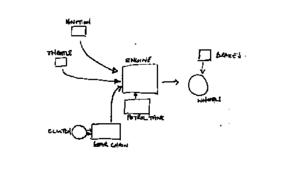
Hand-drawn motorcycle schematic by a study participant Image source: Blackwell, A. F. Correction: A Picture Is Worth 84.1 Words. In Proceedings of the First ESP Student Workshop, pp. 15–22, Fig. 3
Strictly speaking, this experiment wasn't rigorous. But it did make one thing vividly clear: images are easier to understand than text.
The human eye sees pixels; the brain sees structure. The advantage of diagrams is that they bypass the complex abstract reasoning of language and deliver structure directly, making comprehension a dimensionality-reduction process.
But this ability has limits. Humans can only directly perceive three-dimensional space, while biological data often spans thousands of dimensions. A single cell, for instance, might simultaneously record the expression of tens of thousands of genes — stacked together, far beyond human intuitive perception. In the past, we couldn't see clearly. Now AI fills this gap.
High-dimensional data may seem scattered across tens of thousands of dimensions, but because many genes are co-expressed in groups, they tend to cluster near structures of much lower dimensionality. Like a crumpled sheet of paper: it appears to occupy three-dimensional space, but unfolded, it's still a flat plane. AI can flatten these tangled, folded structures, extract key features, and capture the complex structures of the real world.
In the AI era, humanity's ability to see has once again broken through the boundaries of sensory perception across temporal and spatial scales. For drug development, this is especially critical. Because if you look back at every leap in pharmaceutical history, you'll find that almost all of them began with new ways of seeing.

Before 1850, pharmaceutical science didn't yet exist; medical knowledge came mainly from experience. People knew certain herbs, animal products, and minerals might be effective, but couldn't explain why.
As organic chemistry and natural product purification techniques matured, drugs began to enter research view as defined chemical molecules — humanity saw the drugs themselves. In 1897, Bayer researchers prepared acetylsalicylic acid for medicinal use, marking an important milestone in this phase.
Meanwhile, the development of microbiology gradually enabled understanding of the causes of certain infectious diseases. In 1928, Fleming discovered penicillin; through subsequent development, it became a major breakthrough in anti-infective therapy. This research path also clarified the connection between disease etiology and drug action.
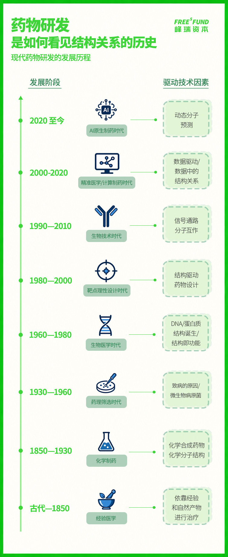
The parallel development of scientific pharmaceutical history and structural biology history
In modern times, pharmaceutical research began intersecting more closely with molecular biology and structural biology. In 1953, the DNA double helix was discovered; in 1958, a low-resolution three-dimensional structural model of myoglobin was published. These discoveries allowed researchers, for the first time, to understand the forms and functions of life molecules from a three-dimensional structural perspective.
As X-ray crystallography, nuclear magnetic resonance, and other technologies gradually matured, we became able to resolve the structures of more and more biological molecules, and structure-based and target-based drug design gradually developed.
Entering the 21st century, high-throughput experimentation and omics technologies advanced rapidly, and researchers began acquiring increasing amounts of data about signaling pathways and molecular interactions. The horizon of drug development expanded from single targets and static structures toward complex networks of molecular relationships. Advances in computational methods and AI also helped researchers begin attempting to predict from these data how molecules move and interact. Drug development thus started moving toward the era of AI-native pharmaceutical research.
Every additional thing humanity sees adds another degree of understanding and intervention space for drug development. But has the living system become sufficiently transparent? The answer: not yet.
In our pharmaceutical history, you can see that many key advances have occurred in the spatial dimension. X-ray crystallography, cryo-EM, and other technologies help researchers see molecular forms, but what they capture is mostly a static snapshot at a particular moment. On the temporal scale, we still lack effective means to continuously track change. How one state transforms into another, in what sequence changes occur, and how they propagate through molecular and cellular networks — much of this remains blank.
In other words, we can see the starting point and the endpoint, but not the middle. What actually happens between a drug entering the human body and ultimately taking effect remains a "black box." This "black box" is the intermediate state of living systems.

What we call the intermediate state today is a generalization of states and transformation processes between initial and final states, across different scales, that have not yet been sufficiently observed.
Intermediate states may appear as transient protein conformations, complexes formed during molecular binding, or state transitions of cells after perturbation.
People often say large models are like black boxes — we can only guess how they operate from their inputs and outputs. Living systems face a similar black box: researchers can record its state at different time points, but struggle to fully see how these states transform continuously.
The difficulty of observation mainly stems from the vast spans of living systems in time and space: atomic motion may occur within femtoseconds, protein conformational changes may last from microseconds to seconds, and cellular responses may take hours; drug action begins at the microscopic scale, while the resulting therapeutic effects and disease changes manifest at the levels of cells, organs, and the whole body. Moreover, data across different scales are difficult to align with each other, and some observation methods may alter the system's original state, further increasing the difficulty of state reconstruction.
When key intermediate processes lack observation and explanation, even if researchers know the results before and after intervention, it's hard to pinpoint where the action begins, which step is blocked, or which mechanisms determine the effect.
So the clearer our understanding of intermediate processes, the more likely drug design can shift from outcome screening to process inference: first judging which pathways a drug might pass through and which key nodes it might affect, then designing molecules, arranging experiments, and revising plans according to the trajectory of change. This way, drug development can reduce some ineffective trial and error, improve design and verification efficiency, and increase the probability of candidate drugs successfully advancing.
In the past, researchers used various experiments and simulations to try to inch closer to those missing intermediate processes. But as mentioned above, this hasn't been easy.
So today, can we accomplish this with AI?
The answer is yes.

AI provides new methods for seeing intermediate states.
Models combine experimental data with applicable biological or physicochemical constraints to reconstruct, simulate, and predict missing life processes between observations, bridging microsecond-scale molecular dynamics data with month- to year-scale clinical observations across scales. We summarize this path as "dynamic visualization."
For example, a movie consists of frame-by-frame still images; when adjacent frames are connected, the viewer perceives motion. The medical equivalent is that experiments provide limited "frames," and models infer the changes that might occur between frames.
If traditional structural biology mainly answered "what does it look like," dynamic visualization answers "how does it become this way." When AI fills in the missing intermediate states between observations, researchers have the opportunity to trace causal relationships between preceding and following states along temporal clues, gradually turning living systems from "black boxes" into "white boxes."
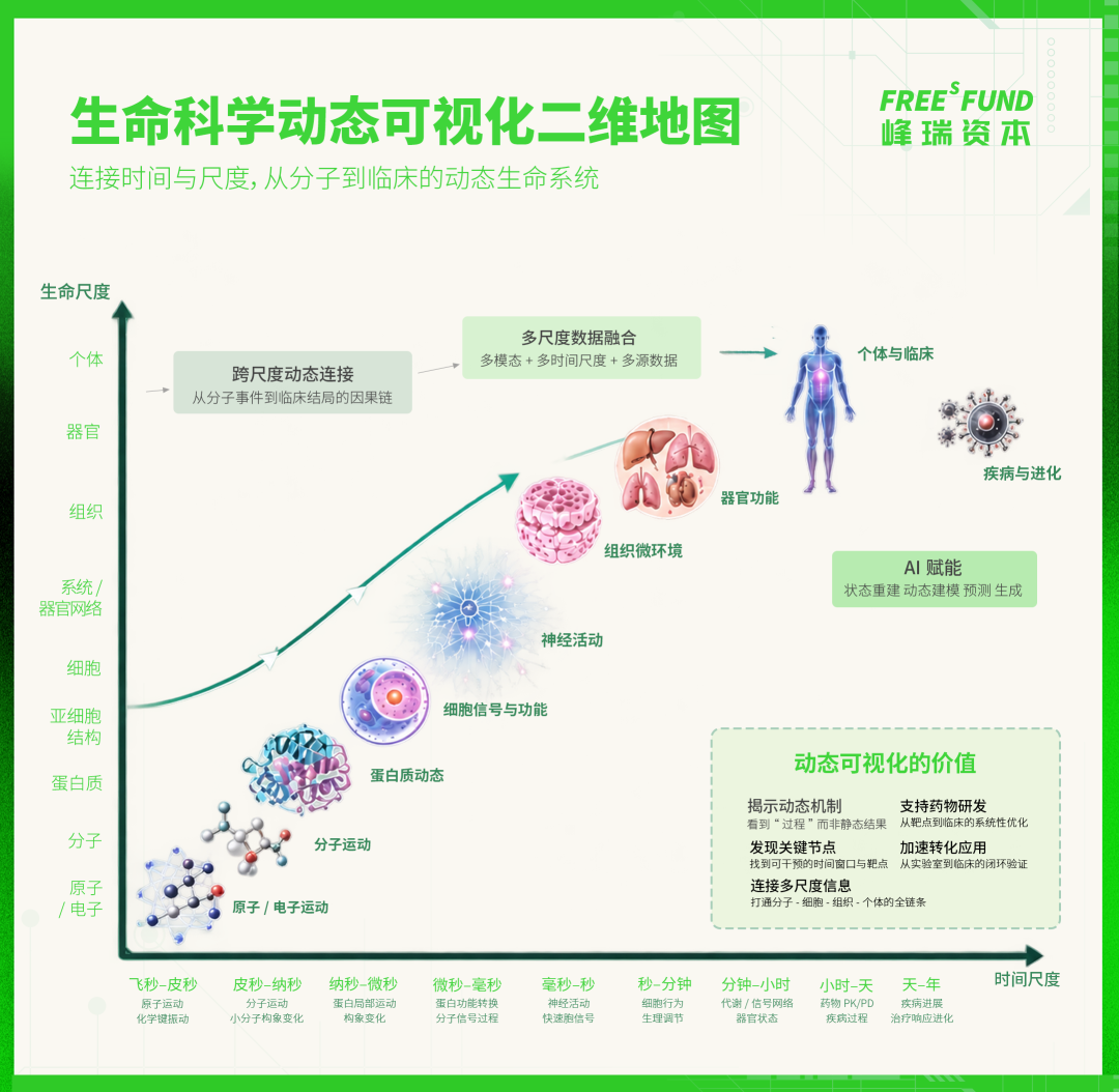
Returning to concrete R&D, we've selected three fascinating and representative cases: cyclic peptides formed from amino acid chains that may change shape with their environment, like "molecular chameleons"; virtual cells that simulate cellular reactions in silico to predict intervention outcomes; and molecular glues that act like "glue" to facilitate specific protein-protein interactions.
From understanding, to inference, to design, they provide three points of observation:
- Cyclic peptides illustrate why dynamic information needs to be incorporated into models;
- Virtual cells demonstrate how AI proposes and validates hypotheses about post-perturbation states;
- Molecular glues further explore how dynamic relationships can become objects of drug design.
Together, the three cases point to one transformation: intermediate processes that were once difficult to observe are gradually entering computable, verifiable R&D frameworks.

In 2026, the cyclic peptide field saw two landmark developments:
- Johnson & Johnson's icotrokinra hydrochloride tablets were approved in the United States and China, becoming the first oral cyclic peptide drug targeting the IL-23 receptor;
- Merck & Co.'s enlicitide received FDA approval, becoming the first oral PCSK9 inhibitor approved by the FDA.
The two approvals demonstrate that oral cyclic peptides are further expanding their application boundaries, providing real-world context for conformational ensemble research (the multiple spatial conformations a molecule may adopt in a given environment and their distribution). Cyclic peptides typically have relatively large molecular weights, with numerous amide bonds, hydrogen bond donors and acceptors, and large polar surface areas. By traditional small-molecule experience rules, these characteristics would pose difficulties for oral absorption.
That some cyclic peptides can overcome the barrier of oral absorption relates to their ability to switch conformations with their environment. Cyclosporin A is a classic example: in aqueous environments, it exposes some polar groups; when approaching a nonpolar environment, it collapses into a more compact conformation, hiding polarity through intramolecular hydrogen bonds; upon reaching the target vicinity, it can switch again to a conformation favorable for binding.
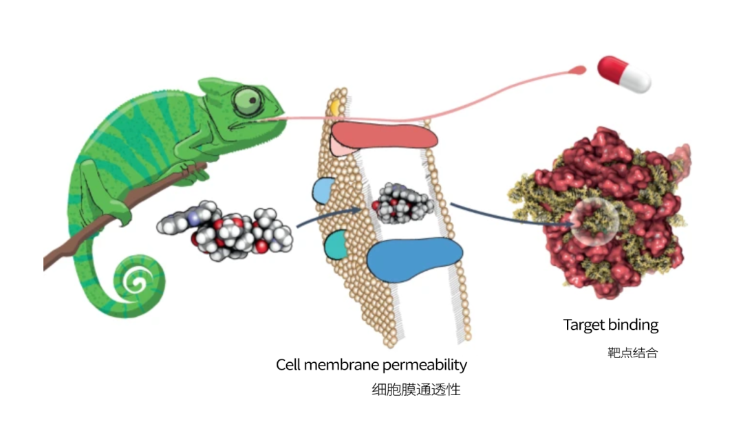
Schematic of molecular chameleon environmental adaptability Image source: Poongavanam, V. et al. Molecular chameleons in drug discovery. Nature Reviews Chemistry 8, 45–60 (2024).
Cyclosporin A and other "molecular chameleons" highlight a variable easily overlooked by static metrics: a molecule's ability to switch conformations with its environment. This means that whether a cyclic peptide can cross the cell membrane cannot be determined solely by polar surface area under a single conformation; we also need to observe how it changes conformation in different environments.
The EnsembleCycPerm model proposed by the Vial algorithm team follows this line of thinking, integrating cyclic peptide sequence, physicochemical properties, and multiple possible conformations in aqueous and chloroform environments to predict passive permeability in the Parallel Artificial Membrane Permeability Assay (PAMPA).
The model focuses not just on how large a cyclic peptide's polar surface area is, but on how much it can change across environments. The researchers describe this change using the difference in three-dimensional polar surface area between aqueous and chloroform environments (ΔPSA3D). In the model's SHAP analysis, this was the single most contributory feature to permeability prediction. In other words, beyond seeing how much polarity a molecule exposes, we need to see whether it can "retract polarity" as the environment changes.
This provides a path toward dynamic visualization for cyclic peptide design. EnsembleCycPerm incorporates conformational ensembles across environments and their changes into permeability prediction, offering reference for candidate molecule screening and lead optimization.

A cyclic peptide's conformational change occurs within the molecule itself. Expanding the view to the cell, researchers face a network pulled by numerous signals in concert.
Traditional research uses gene knockout, drug treatment, or other molecular perturbations to observe subsequent cellular changes. One experiment can establish a relatively clear "perturbation—outcome" relationship; accumulated experiments gradually piece together cell signaling pathways. But faced with complex signaling networks, experiments can still only cover limited relationships, with many interactions and dynamic changes remaining outside observation.
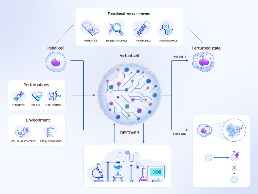
"Predict—Explain—Discover" framework schematic for virtual cells Image source: Noutahi, E. et al. Virtual Cells: Predict, Explain, Discover. arXiv:2505.14613 (2025).
Virtual cells, as learned simulators, use data from real cells under different genotypes, drug treatments, gene edits, and environmental conditions to learn how cell states change. Once the model is established, researchers can perform large numbers of "what if" virtual perturbations in silico, inferring possible cellular responses and dynamic processes and interaction relationships in signaling networks not yet covered by experiments.
Their significance for AI pharma lies in connecting models and experiments into a closed loop: real cell data trains the virtual cell; the model infers changes in cell state through virtual perturbations, presenting prediction results through dynamic visualization; these predictions then return to real experiments for validation, and new experimental data continues to refine the model. In this cycle, the processes between perturbation and outcome begin entering a computable, verifiable dynamic visualization framework.
As mentioned earlier, research on temporal relationships has long been a weak point for humans. What virtual cells attempt is precisely to fill this gap, which is also an important reason they have attracted significant attention in the industry.
Baiyao Technology, a FreeS Fund portfolio company, had its core founding team explore this path relatively early. Since 2023, the team has continuously invested in related research, making it one of the earlier domestic teams to explore AIVC technology. Building on this foundation, Baiyao Technology launched its virtual cell foundation model CellOS after its founding.
CellOS is trained on 390.5 million human single-cell transcriptome data points, with 12 billion parameters, and innovatively adopts the LLM-JEPA architecture. The model learns stable representations of living systems from two perspectives — gene expression and cell state — to understand cell states and predict responses under perturbations such as gene editing, drug action, and environmental changes. Baiyao is committed to building a full-stack AIVC platform, enabling research processes that originally relied on extensive trial and error to gradually shift toward a paradigm leap of "AI simulation—experiment synergy": researchers can first conduct large-scale simulations in virtual environments, comparing possible state changes from different interventions, then hand high-value candidates to real experiments for testing; experimental results feed back into the model, forming a closed loop of "simulate—predict—verify—learn."
What virtual cells solve is fundamentally a spatiotemporal dynamic visualization problem. Starting from perturbation data, they propose predictions for cellular responses not covered by experiments and provide clues for potential regulatory relationships. Only as models and experiments are repeatedly calibrated can scattered observations gradually connect into more complete cellular signaling networks.

Virtual cells demonstrate the basic path of state inference. With molecular glues, the question advances further: how does a protein interface that was originally difficult to form emerge with the participation of a small molecule, and persist long enough to trigger ubiquitination and protein degradation? Dynamic relationships thus directly become objects of drug design.
In the targeted protein degradation field, PROTACs (proteolysis-targeting chimeras) use a bifunctional molecule — one end binding the target protein, the other binding an E3 ubiquitin ligase, connected by a linker that pulls the two together. If the resulting complex can support effective ubiquitination, the target protein can be tagged with ubiquitin and degraded by the proteasome. This design path is relatively clear, but the molecules are typically large, posing druggability challenges.
Molecular glues don't rely on such linkers; instead, they facilitate binding between target proteins and E3 ligases through small molecules. Taking CRBN-related molecular glues as an example, CRBN is the receptor in the E3 ligase complex responsible for recognizing substrates. The ternary complex formed by small molecule, CRBN, and target protein can further promote target protein ubiquitination and degradation.
The difficulty concentrates on the formation process of the ternary complex: how the small molecule creates a new recognition interface, which proteins can match with it, and how the three form a stable structure — difficult to judge from binding results alone.
As structural biology, proteomics, and chemical screening accumulate more data, AI is beginning to have the opportunity to connect these scattered observations: finding potentially recruitable new substrates from protein structures, predicting potential interactions, and handing them to experiments for validation.
Monte Rosa Therapeutics' QuEEN platform (Quantitative and Engineered Elimination of Neosubstrates) represents one current-stage attempt. It combines artificial intelligence, chemical libraries, structural biology, and proteomics to improve new substrate discovery efficiency, pushing molecular glue R&D toward a "model—screen—verify—optimize" process.
We judge that molecular glue R&D will continue moving toward dynamic visualization.
Models need to further describe how interfaces change after small molecule binding, how target proteins approach each other, and how ternary complexes form and maintain stability. When this process can be reconstructed, predicted, and verified, rational design of molecular glues will gain a more complete foundation.
For cyclic peptides, models incorporate conformational ensembles across environments into permeability prediction; for virtual cells, perturbation data is being used to predict unobserved cellular responses; for molecular glues, dynamic modeling of ternary complex formation still points to the next development direction. Together, the three cases indicate that AI pharma is beginning to move toward dynamic visualization.

Returning to the early-stage investment perspective, the development of dynamic visualization is a critical path. FreeS Fund will continue to track opportunities emerging in the AI-native pharmaceutical field, with particular attention to wet-dry closed-loop experimental platforms that leverage AI to address traditionally undruggable or difficult-to-drug targets.
From relying on empirical drug use, to using AlphaFold to predict protein structures, humanity's understanding of living systems continues to deepen. Now, the new combination of seeing + AI will once again expand the boundaries of human ability to understand life.
Repeated verification allows predictions to settle into knowledge, while new questions and intervention paths often come from researchers' intuition about complex relationships. Poincaré once said: "We prove by logic, but we invent by intuition."
Intuition is not the opposite of logic. Often, it comes from long-term observation and compression of structure. When AI can transform invisible intermediate states into observable, challengeable, and verifiable dynamic models, researchers' intuition will also gain new material.